
Image of the structure of the mouse Hippocampus (Image courtesy of http://www.gensat.org).
How are new memories created?
This is a fascinating question in neuroscience and at the very core of what makes us human. After all, our entire concept of ourselves is defined by our memories and without them, are we even ourselves? This is a pretty lofty philosophical discussion… but today we’re only interested in the neuroscience of memory.
In specific, what happens to individual neurons in the human brain when a new memory is created and recalled?
Researchers at the University of California-Los Angeles performed a study in humans that has shed some light on this important question. Published recently in the journal Neuron, the novelty of the study involved recording how many times a neuron would fire during a specially designed memory test. In other words, the scientists were able to monitor what happened to individual neurons in a human being as a new memory was being created!
This article is open access (able to downloaded and distributed for free). The article can be found here or download the pdf.
Before I go into what the researchers found, let’s see how it was done.
The subjects in the study were patients being treated for epilepsy. As part of their clinical diagnosis, they had been implanted with an electrode, a tool used to measure neuronal activity or in other words, the electrode measures how often a neuron fires. The fact these patients already had an electrode inserted into the brain for clinical reasons made it convenient for the researchers to conduct this study.

Left Temporal Lobe (www.wikipedia.org)
The brain region in which the electrode was implanted is called the medial temporal lobe (MTL). The image to the right is of the left human temporal lobe. The medial region of the temporal lobe is located more towards the center of the brain.

Human Hippocampus (www.wikipedia.org)
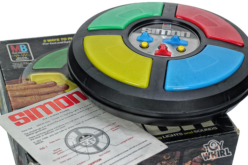
One specific region of the MTL, the hippocampus, is believed to be the primary brain region where memories are “stored”. Specifically, previous studies in animals and humans have suggested that the MTL and hippocampus are very important to encoding episodic memory. Episodic memory involves memories about specific events or places. In this study, the example of episodic memory being used is remembering seeing a person at a particular place. Another example: the game Simon™ can be considered a test of your brain’s ability to rapidly create and recall short-term episodic memories!
*Note: Episodic memory is considered one of the main bifurcations of declarative memory, or memories that can be consciously recalled. The other type of declarative memory is semantic memory, which are memories of non-physical/tangible things, like facts.
To test the episodic memory of remembering a person at a particular place, images were presented to the patients while the neurons were being recorded. There were 5 different tasks (all completed within 25-30min). See Figure 1 below from the paper.
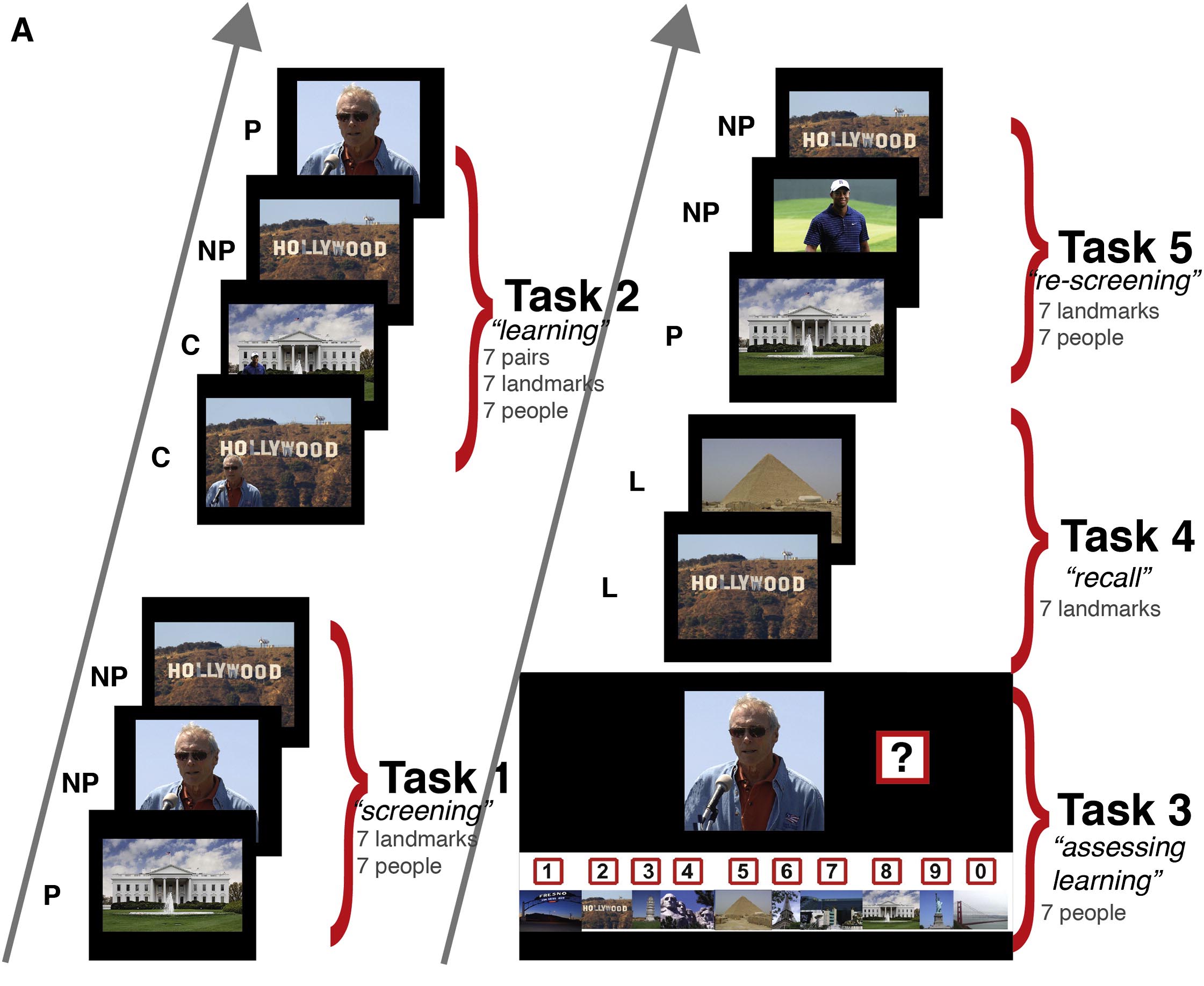
Figure 1: Experimental Design
First, a pre-screening was done in which the patients was shown many random images of people and places. The activity of multiple neurons was recorded and the data was quickly analyzed then 3-8 pairs of images were compiled. In each pair, 1 image was “preferred” or “P” image, meaning the neurons being recorded fired when the “P” image was shown. The second image was “non-preferred” or “NP” image, meaning the neurons did not respond to it when it was shown.
The first task is the “Screening” test. Each “P” and “NP” image was shown individually and the neurons response to each was recorded. As you would expect, the neuron would fire heavily to the “P” image and not very much to the “NP” image.
The second task was the “learning task” in which a composite image of each of the “P” and “NP” image pairs was made. The person in the “P” image was digitally extracted and placed in front of the landmark in the “NP” image. After the composite images were shown, the individual images were shown again.
For example, in one image pair for one patient, the “P” image was a member of the patient’s family while the “NP” image was the Eiffel Tower (for this example, see Figure 2). The composite image in the “learning” task was the family member in front of the Eiffel Tower. Another example of a “P” image was Clint Eastwood and the “NP” image was the Hollywood sign. The composite image would therefore be Clint Eastwood in front of the Hollywood sign. (However, in some image pairs the “P” image was a place and “NP” image was a person).
The third task was “assessing learning”. The image of just the person in the composite image was shown and the patient had to pick out the correct landmark he/she was paired with. For example, the picture of the family member was shown and the patient would have to pick out the Eiffel Tower image.
The fourth task was the “recall” task. The landmark image was shown and the patient had to remember and say the person it was paired with. For example, the Eiffel Tower was shown and the patient had to say the family member’s name.
Finally, the fifth task was a “re-screening” in which each individual image was shown again so the neuron’s activity could be compared to the Task 1, pre-learning.
The activity of multiple neurons were recorded for each image for each of the tasks. The data was then analyzed in number of different ways and the activity of different neurons was reported.
And what was found?

Figure 2: Response of Neurons in the Hippocampus from a Patient
Let’s go back to the family member/Eiffel tower example. The researchers were able to show that a neuron in the hippocampus responded heavily to the picture of the family member (“P” image) but not to the Eiffel Tower (“NP” image). After showing the composite image, the neuron now responded to the Eiffel Tower too in addition to the family member! (The neuron also fired a comparable amount to the individual family member image as the composite image).
As you can see in Figure 2, each little red or blue line indicates when a neuron fired. For example, in Task 1 you can clearly see more firing (more lines) to the “P” image than the “NP” image. You can see that after Task 2, the neuron responds to either the “P” or “NP” image (especially obvious in the Task 5). The middle graph indicates the firing rate of the neurons to the “NP” image and it clearly shows increased firing rate of the neuron after learning (AL) compared to before learning (BL). It may look small, but the scientists calculated a 230% increase in firing rate of the neuron to “NP” image after the learning/memory task took place!
What does this mean? It means that a new episodic memory has been created and a single neuron is now firing in a new pattern in order to help encode the new memory!
This was confirmed the other way around too. In another patient, the “P” neuron was the White House and the “NP” image was beach volleyball player Kerry Walsh. The neuron that was being recorded fired a lot when the image of the White House was shown but not so much for the Kerri Walsh image. Then the composite image was shown and the learning/recall tasks were performed. The neuron was shown to fire to both the White House image AND the Kerry Walsh image! The neuron was responding to the new association memory that was created!
Keep in mind these are just two examples. The scientists actually recorded from ~600 neurons in several different brain regions besides the hippocampus but they only used about 50 of them that responded to visual presentation of the “P” image, either a person or a landmark (the identification of visually responsive neurons was crucial part of the experiment). Remarkably, when the firing rates of all these neurons was averaged before and after the memory/learning tasks, a similar finding to the above examples was found: the neuron now responded to the “NP” image after the composite was shown!
Many other statistical analyses of the data was done to prove this was not just a fluke of one or two neurons but was consistent observation amongst all the neurons studied but I won’t go into those details now.
But what’s going on here? Are the neurons that respond to the “P” stimulus now directly responding to the “NP” image or is more indirect, some other neuron is responding to the “NP” which in turn signals to the “P” neuron to increase in firing? The authors performed some interesting analyses that both of these mechanisms may apply but for different neurons.
Finally, were all the recorded neurons that were engaged in encoding the new episodic memory located in the hippocampus? The answer is no. Responsive neurons were identified in several brain regions besides the hippocampus including the entorhinal cortex and the amygdala. But most of the responsive cells were located within the parahippocampal cortex, a region of the cortex that surrounds the hippocampus, thus not surprising it is very involved in encoding a new memory.
In conclusion, the scientists were able to observe for the first time the creation of a new memory in the human brain at the level of a single neuron. This is an important development but such a detailed analysis has never before been done in humans and, most importantly, in real time. Meaning, the experiment was able to observe the actual inception of a new memory at the neuronal level.
However, one major limitation is that the activity of these neurons were not studied in the long term so it’s unknown if the rapid change in activity is a short-term response to the association of the two images or if it really represents a long-term memory. The authors acknowledge this limitation but the problem is really in the difficulty of doing such studies in humans. It’s not really ethical to leave an electrode in someone’s brain just so that you can test them every week!
But what does all of this mean? The authors do suggest that the work may help to resolve a debate that has been going in on the psychology field since the 40s. Do associations form gradually or rapidly? These results strongly suggest new neurons rapidly respond to encode the new memory formation.
But how will these results shape the neuroscience of memory? The answer is I don’t know and no one does. Thus is the rich tapestry of neuroscience, another thread weaved by the continuing work of scientists all over the world in order to understand what it is that makes us human: our brains.
