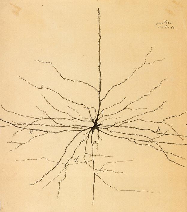
A single, pyramidal neuron. Illustration by Ramon Y Cajal S., ca. 1899.
The image above is a hand drawing of a single neuron (specifically, a pyramidal neuron) completed by the brilliant Spanish scientist Santiago Ramon y Cajal (1852-1934): the father of modern neuroscience. He made many remarkable preparations (slides containing intact cells from brains) that he used to make countless impressive drawings of brain cells, or neurons. We have learned a wealth of information about the composition of the brain and the structures of neurons from the work of Ramon y Cajal. His images of neurons are so detailed, accurate, and just plain beautiful, they are still used in universities and labs throughout the world today.
Like discussed in the previous post, the brain is the organ that drugs act on to change the behaviors and thoughts of the drug user. Specifically, drugs act on neurons and today’s post will be an introduction to the basic biology of these elegant cells.
A Quick Note: This and future posts are intended to be modular, meaning that each post should be an independent parcel of knowledge. If you already know about (or are not particularly interested) in the topics covered in the post, skip it and read a different one. This post is largely a basic neuroscience one and many of the other posts may speak about science/neuroscience more generally but I will try to tie most posts into the general theme of drug addiction as a biological/medical disease and/or the brain vs mind theme.
As I was saying: like every other organ in the body, the brain is made up of cells. As many of you know, the cell is the basic unit of biology. This concept is also known more generally as the Cell Theory of Biology. Basically, an organ’s function is derived from its component cells. The heart beats because of the coordinated efforts of the heart cells (cardiomyocytes) that the heart is made of and the liver functions because it is made up of many types of liver cells (hepatocytes) that all have different jobs to do. And the brain works because of the efforts of brain cells.

Neurons (in green) and Glial Cells (in red) in the Cortex of the Mouse Brain. Image courtesy of Ana Milosevic, The Rockefeller University.
There are two main types of brain cells: neurons and glial cells. Neurons are the real workhorses of the brain while glial cells are considered support cells that help maintain brain structure and “take care” of neurons. We will not discuss glial cells at all today but will focus exclusively on neurons.
The human brain literally contains billions of neurons and these billions of neurons make trillions of connections to one another. The amazing thing is that many of these neurons operate in the same basic way: they receive a signal from a different neuron, they process the signal, and then they conduct and transmit that signal to different neurons. The details about what signals, which and how many neurons are communicated to/with, and the consequences of those signals is highly specific to the individual types of neurons and neuronal populations in the brain and is extremely complicated.
But this very general function: listen-process-talk (or stay quiet in some cases) forms the basis for everything that your brain does.
The thing I find the most extraordinary about neuroscience is all your feelings, thoughts, desires, actions, and everything you know or will ever know is because of neurons communicating with one another. And drugs have the potential to drastically change this communication (more to come).
Every animal on the planet has neurons and, once again, the basic function of human neurons, and monkey neurons, and bird neurons, and even insect neurons is the same thing: receive and conduct signals (listen-process-talk). Just as a quick-and-dirty comparison, it’s estimated that the human brain contains about 100 billion neurons while the nematode worm C. elegans (a tiny worm used for study in many labs) has only 302 neurons.
So I’ve mentioned the neurons receive and transmit signals but what signals am I talking about? Electrical and chemical signals. Each neuron has the potential to fire or to not fire. When a neuron fires, it literally generates an electrical charge and moves that charge along its length (the neuron conducts an electrical signal). When the electrical activity reaches the end of the neuron, the electrical signal is then translated into a chemical signal, which is released onto another neuron. The neuron that receives the chemical signal is then able to translate it back into electricity and fire again, thus passing the electrical signal onto another neuron and so on and so forth (and in some cases, the chemical signal says “STOP” and no electricity is generated).
The above paragraph is very complicated and will be discussed in more detail in this post and the next few. But for now let’s just take a look at the structure of the neuron in more detail. Like everything in biology, the structure (how it looks or is arranged) of something tells you a great deal about its function.

Figure 1: Diagram of a Neuron.
Figure 1 is a simple diagram of a neuron that I made.
You will notice two main parts of the neuron: a head with many branching structures and then a long tail with more branches. This head part is called the cell body or soma. Like other cells, this is where the organelles (mitochondria, ribosomes, endoplasmic reticulum…if these don’t sound familiar, do a quick search for “cell biology”) are located and where the normal cell maintenance activities occur (same as every other cell). This is also where all the signals received by the neuron are processed. And like most plant and animal cells in nature, neurons have a nucleus, the control center of the cell and the region that contains the cell’s DNA (I may do a primer on some basic molecular biology at some point).
At the base of the cell body is a long tail-like structure and this is one of the most important features of the neuron: the axon. The axon is how the electrical signals get transmitted to other neurons. Some axons are incredibly long. For example, motor neurons (neurons that receive signals from the brain in order to tell your muscles to contract) extend the entire length of your body! Just think about that for a second and then move your finger or toe. Any lag? Probably not, the motion is almost instantaneous and that’s because the axon is really, really good at conducting electricity!

Figure 2: The Myelin Sheath. A cross section of an axon surrounded by a Schwann cell, which forms the protective myelin sheath around it.
To help it do this, the axon is covered in a fatty substance called myelin (MY-elin). Myelin forms a tight bundle around the axons called the myelin sheath. Figure 2 is a cross section of the axon. The myelin is actually part of a special type of cell called a Schwann cell, and these cells quite literally are wrapped around axons to form the myelin sheath (Figure 1 and 2). Myelin is so important because it acts like an insulator, just like an extension chord or other electrical wires are covered in rubber or some other material. The insulating effect of myelin (just like in other wires) helps to maintain and conserve the electrical signal.
Note that there are gaps between the Schwann cells (gaps in the myelin sheath) along the length of the axon (Figure 1). These are called the nodes of Ranvier. They play an important role in conducting the electrical signal but we’ll save this discussion for later.
Finally, at the end of the axon is a series of branching structures called the axon terminal (Figure 1). This is where the electrical signal is translated into a chemical signal. That is to say, once the electrical signal reaches the axon terminal, it causes little packets of very special signaling chemicals, called neurotransmitters, to be released from the axon terminal. We’ll spend a lot of time talking about neurotransmitters (especially dopamine, the neurotransmitter that’s very important in understanding addiction).
Now let’s go back up to the cell body. You’ll note that there are many branching structures extending from the cell body. These are called dendrites. Also note that other neurons are projecting onto the dendrites. These projections are actually axon terminals from different neurons. When a neurotransmitter is released from an axon terminal, it acts on a dendrite. This junction of one neuron’s axon terminal with another neuron’s dendrite is the called the synapse.
The synapse is not a physical connection but a very tight space between neurons. This is important because it is in this empty space that the neurotransmitters are released. Understanding the function of the synapse is crucial for understanding anything in neuroscience and when we discuss neurotransmitters we’ll talk more about the synapse.
So just to return to our listen-process-talk analogy for a moment, the dendrites would be the ears, the part of the neuron that listens, or receives the signal. The cell body is where the chemical signals are processed and then this may result in the neuron firing. If it fires, the electrical signal is transmitted along the axon, which would be like the vocal chords or tongue, both of which help in the act of talking. Finally, the mouth in our analogy is the axon terminal, the part of the neuron where the “talking” comes from, and the words the mouth “says” are neurotransmitters.

Real Mouse Neurons. Diagram of important neuronal structures. (Modification of original image courtesy of Kinning Poon, Rockefeller University, 2014)
Just to wrap things up, this is an image of real neurons from the mouse brain (hypothalamic neurons if you really want to know). The blue color indicates the nucleus (but don’t worry about the other colors). This image is an immunofluorescent image and it’s a nifty technique to examine detailed cellular structures (yes, I’ll explain this in detail later too. Also added to the list….). I’ve pointed out some of the structures that we talked about today.
Ok, so I guess my goal to write shorter posts is not really happening…
Hopefully you’re not feeling too overwhelmed. Over the course of the next two posts, we’ll dive into how neurotransmitters cause neurons to fire or to not fire.

Pingback: Synapse to it – Dr. Simon Says Science
Pingback: Marijuana has Long-term Effects on the Brains of Adolescents – Dr. Simon Says Science